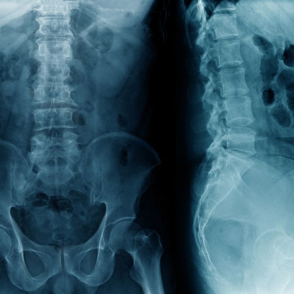
Endoscopic Surgery
Endoscopic Discectomy in Brick Township, NJ
Helping People Relieve Their Pain Related To Their Spine and Return Them to a Higher Quality of Life
Endoscopic Surgery
Open Endoscopically Assisted Tubular Retractor Surgery (Endoscopic Surgery) the least invasive procedure used to treat herniated, protruded, extruded and disc tears that compress or irritate the spinal nerves causing back or leg pain. Patients suffering from many spinal conditions should consider all less invasive options like pain management and endoscopic procedures before ever considering traditional, open or minimally invasive spine techniques including laminectomy, microdiscectomy or spinal fusion. With proper diagnosis and diagnostic injections the minimally invasive physician can pinpoint the cause of the pain and selectively address the painful condition without causing the patient significant post-operative pain or recovery.
Endoscopic Surgery is an outpatient surgical procedure to remove herniated disc material. Using local anesthesia with the help of x-ray fluoroscopy a needle, then guidewire, then dilator and working cannula are placed in succession of each other to open a small 7mm or ¼ inch portal. Once the working portal is established a specially designed endoscope and attached HD camera for magnified video are inserted into the cannula. The physician has superior endoscopic visualization of the anatomy and the herniated disc and with tiny surgical instruments can selectively remove a small portion of the disc that is compressing the spinal nerve and eliminate the patients pain.
The procedure takes about an hour, on average. X-ray exposure is minimal. You normally will feel little, if any pain or discomfort. There are no stitches. Upon completion, the endoscope and cannula are removed and a small Band-Aid is placed over the incision. The amount of nucleus tissue removed varies but the supporting structure of the disc is not affected by the surgery. The access route to the disc consists of only the ¼ inch small puncture site, usually the size of a freckle, in comparison to large incisions required for open surgery.
Open Endoscopically Assisted Tubular Retractor Surgery is different from open lumbar disc surgery because there is no traumatic back muscle dissection, no bone removal, or large skin incision. The risk of complications from scarring, blood loss, infection, and anesthesia that may occur with conventional surgery are drastically reduced or eliminated with this procedure. Open Endoscopically Assisted Tubular Retractor Surgery was invented to be an effective treatment for herniated discs while avoiding these risks.
The Minimally Invasive Advantages to Traditional Spinal Surgery:
- Utilizes a HD camera and endoscope which affords the physician a superior view to that of traditional techniques
- ¼ inch – the smallest incision in spine surgery
- No muscle or tissue tearing thus less scar tissue
- Conscious sedation reduces the risk associated with general anesthesia
- Less post-operative pain and need for narcotic medicines
- Less recovery time needed
- Return to work sooner
What Types of Conditions do Minimally Invasive Surgery Treat?
- Arthritis and Bone Spurs of the Spine
- Bulging Disc
- Discogenic Back Pain
- Herniated Disc
- Failed Back Surgery
- Foraminal Stenosis (Narrowing of the Spinal Canal)
- Sciatica
- Radiculitis or Radiculopathy
- Spondylolisthesis (slipping of the Vertebra)
Types of Minimally Invasive Procedures:
- Transforaminal Open Endoscopically Assisted Tubular Retractor Surgery – treats herniated, bulging, protruded, and extruded discs. Under direct visualization, the physician uses the endoscope to remove any pathology which causes pressure on the affected spinal nerve.
- Endoscopic Assisted Foraminalplasty – treats degenerative disc, foraminal stenosis and facet disease. As the space between the facets diminishes the foramen (natural opening for the spinal and exiting nerves) becomes narrow and begins to compress the nerves. The endoscopic technique uses ronguers, reamers, and small motorized burrs to selectively take some bone in order to enlarge the foramen thus decompressing the nerves freeing them up.
- Endoscopic Assisted Rhizotomy – treats patients suffering from chronic axial back pain. When patients lean forward they are fine but leaning backwards causes significant pain and spasms. Patients who have received some temporary relief from percutaneous medial branch rhizotomy but the pain came back are good candidates for endoscopic rhizotomy. This procedure allows the physician to place a small cannula and endoscope inside the patients back and target visually the medial branch nerve. A radiofrequency probe is used through the endoscope to ablate the medial branch nerve. The results of endoscopic rhizotomy have been significantly better long-term than traditional percutaneous rhizotomy.
Contact Us Today
74 Brick Blvd. Brick, NJ 08723
Please take a moment to tell us a little about yourself and your needs. A member from Jasper Spine Institute will contact you as soon as possible. If your needs are urgent please contact us by phone at (732) 262-0700.